The lesions of HSV have a characteristic appearance and so can be easily diagnosed clinically by naked eye examination.

-
However, in some cases an accurate diagnosis can be obtained by:
- Viral culture of the lesion that shows the definitive presence of herpes virus
- Direct fluorescent antibody (DFA) that shows the presence of specific antibodies to herpes virus
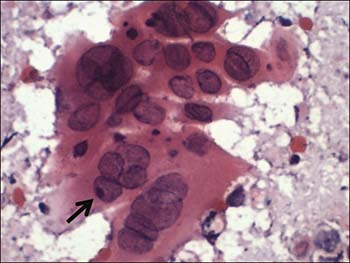
- Tzanck test is a time honored procedure showing presence of specific cells (multinucleated giant cells) on a smear from the herpes lesion
- Punch biopsy for histological examination showing typical cells and characteristic tissue changes
- Examination of cerebrospinal fluid in HSV encephalitis showing typical changes in levels of protein, concentrations of various immunoglobulins, as well as presence of specific antibodies
- Brain biopsy and imaging in cases of HSV encephalitis (inflammation of the brain)


